"A" photos:
(click to enlarge)
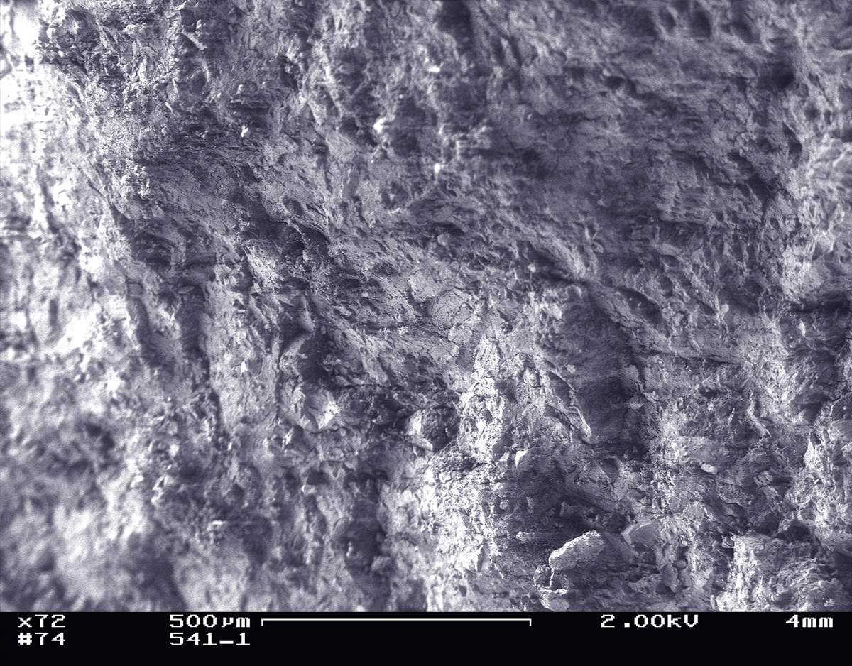
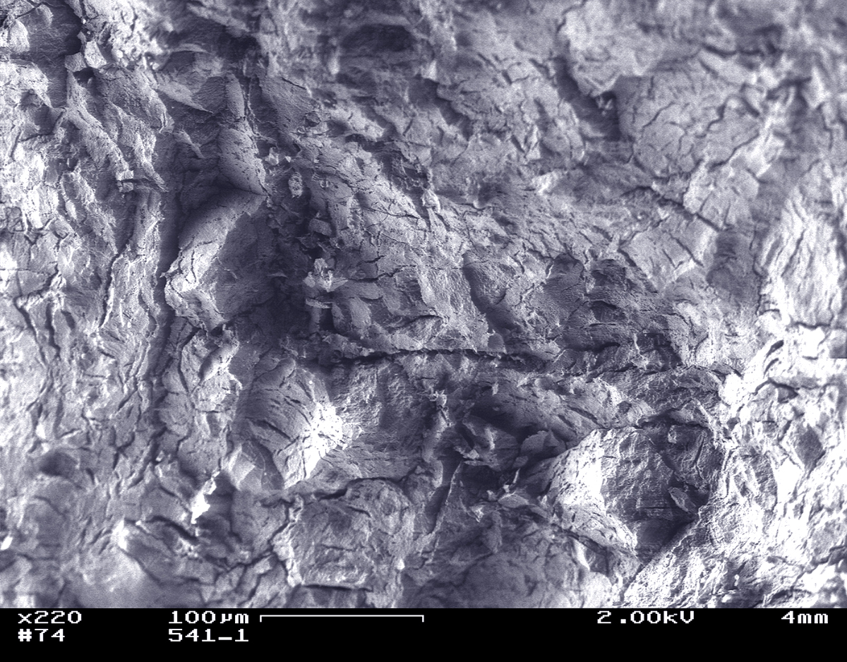
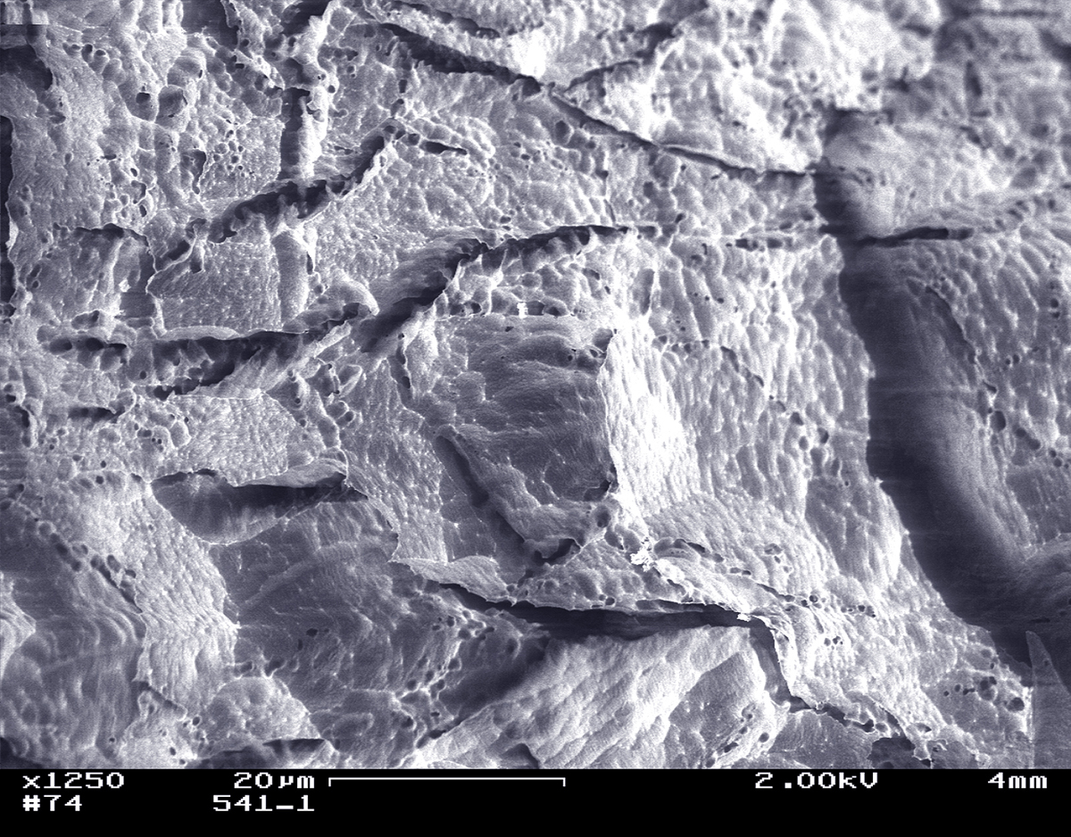
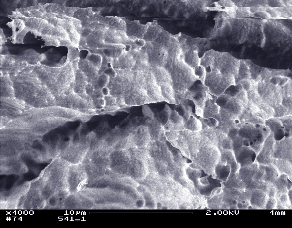
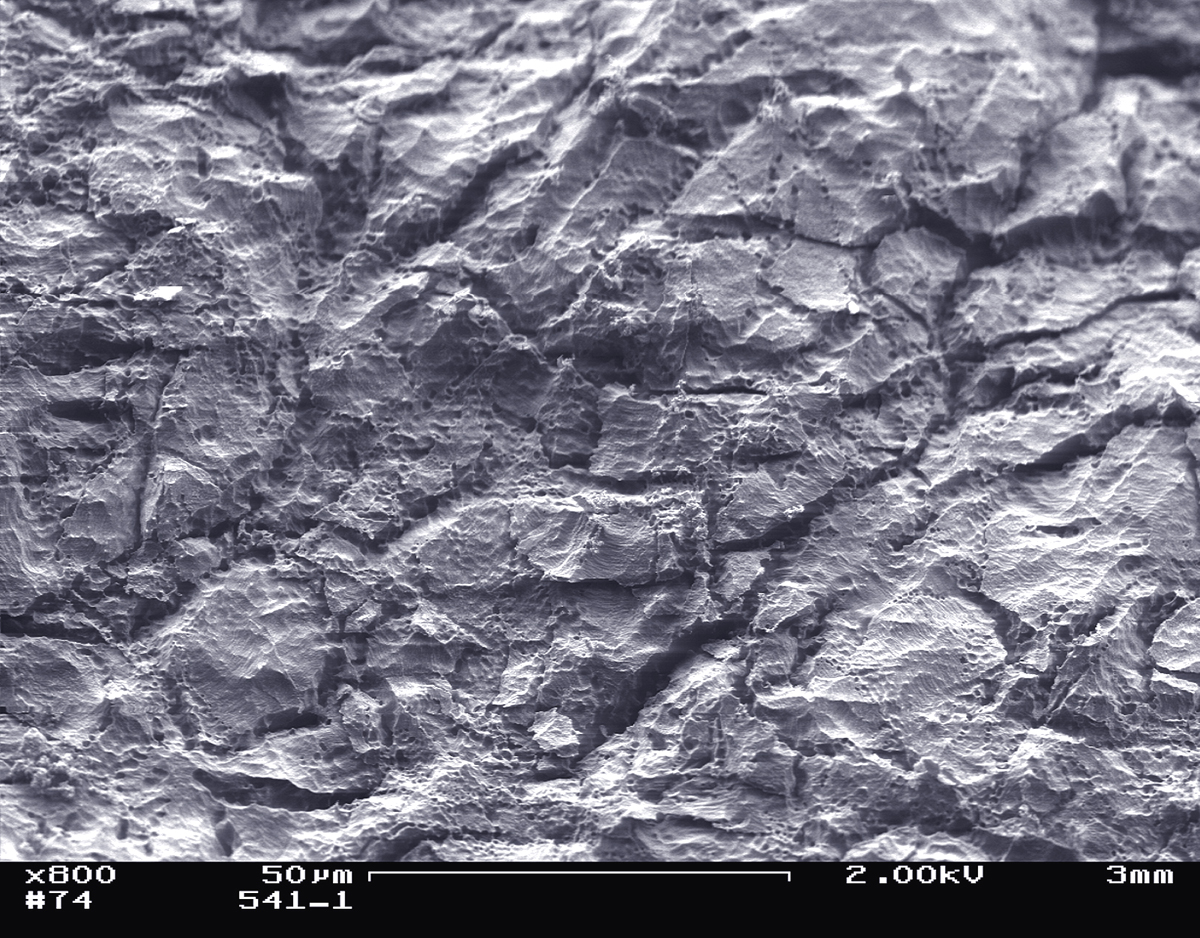
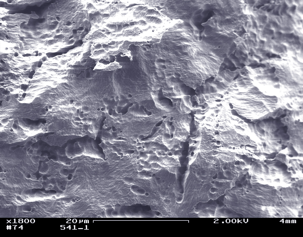
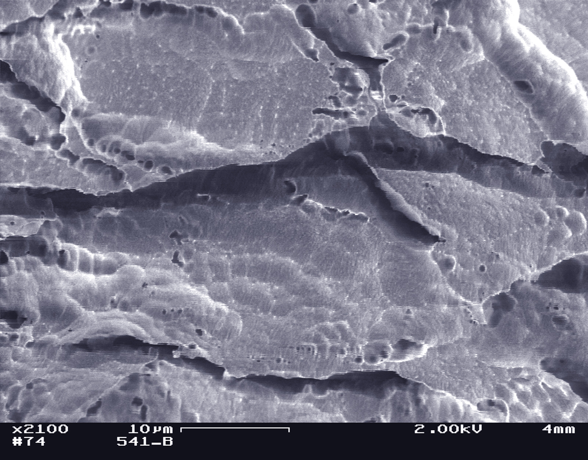
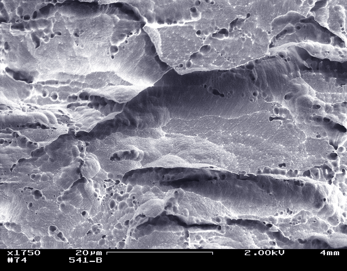
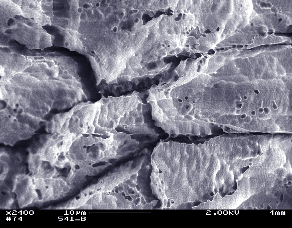
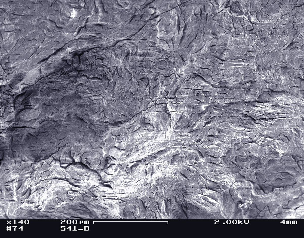
LAS Micrographs
LAS email 6/29/07
Subject: Sample 541
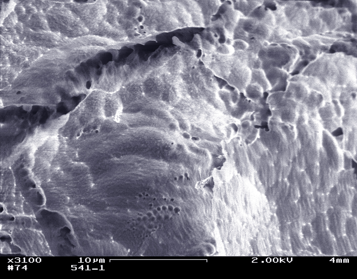
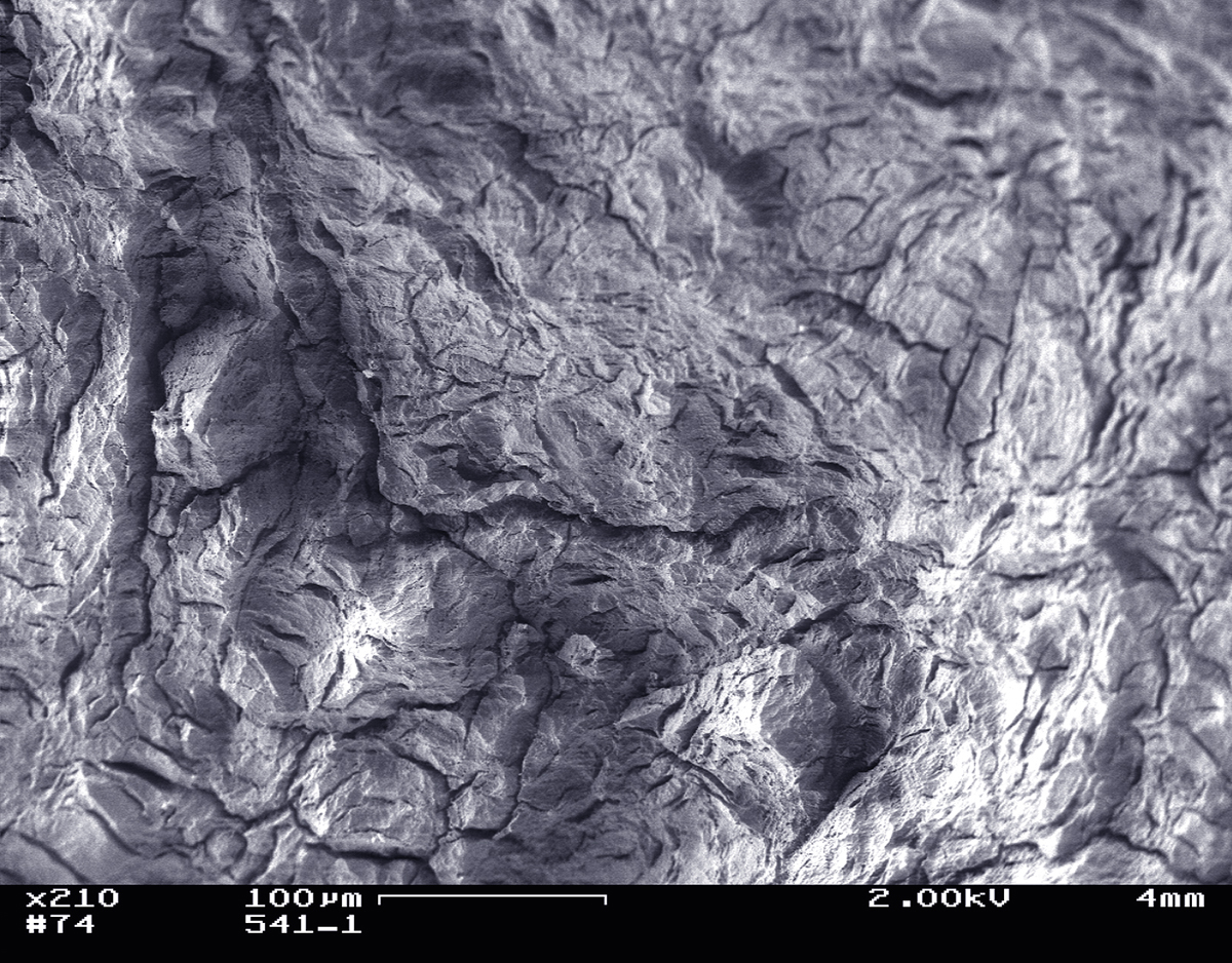
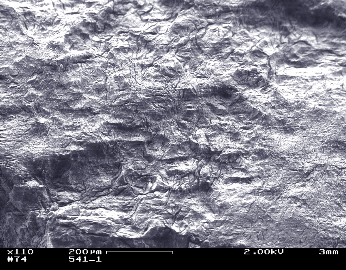
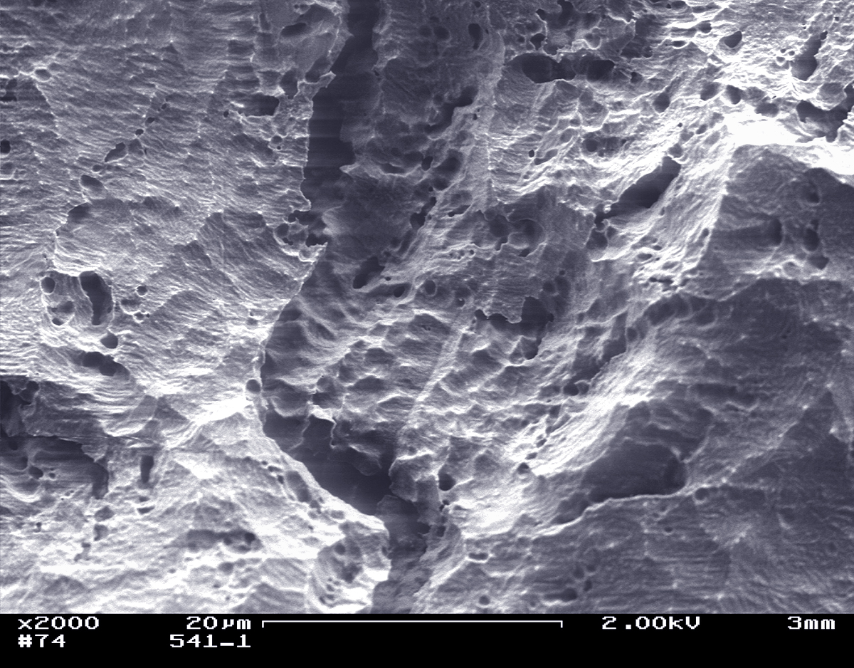
I looked at two different sections of this sample. Cool sample. The fractures continued to open up fairly rapidly, as you can see by comparing A2 to A8, which were taken 21 minutes apart.
Some photos show nice, fine, grain boundaries, others show the deterioration after beam and vacuum exposure. I'll lay these out in the order I took them. In the last 3 photos on this page, note that what may appear as dimples or pits are standing up in relief, not down. (i.e. you're looking at the tops of grains or small grains, not holes.) Sometimes I have to stare at these for a while before the image suddenly "inverts". It's dead obvious when scanning the full surface of the sample, but for some reason can be very hard to see correctly in individual images.
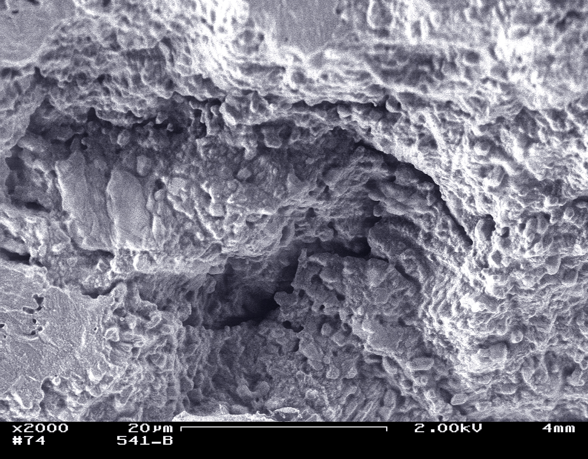
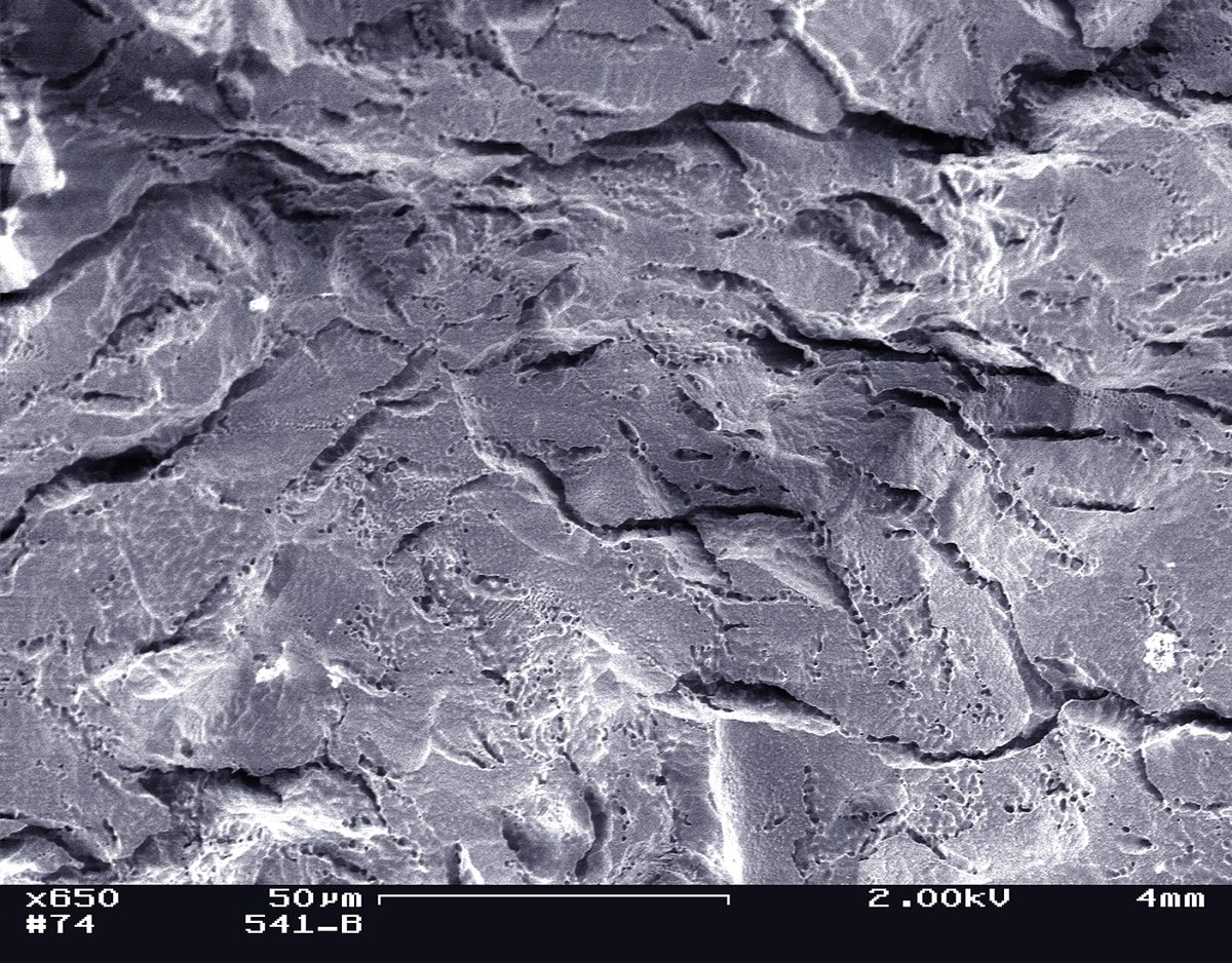
The photos labeles "A" are the first sample, those labeled "B" are the second sample. Good uniformity.
"A" photos: |
(click to enlarge) |
|
|
|
|
|
|
|
|
|
|
|
||
| "B" photos: |
||
|
|
|
|
|
|